Lymph node aspirates in Tuberculosis-Diagnosis: New challenges, new solutions – a second study of 545 patients
Dr. Alexander von Paleske ---- 15.8. 2012 -
Department of Haematology: Alexander von Paleske, MD, FA(Med), FA (Haem / Onc); Owen Chivima, SCMLT
National TB Reference Laboratory: Barbara Murwira, CMLS; Thembela Masuku, SMLS; Tanaka Sakubani, SMLS; Regina Bhebe, SMLS.
Mpilo-Hospital
Teaching Hospital of the University of Zimbabwe
Bulawayo/Zimbabwe
Africa
Introduction
Lymph node tuberculosis is not infrequent in Sub-Saharan Africa. Even though precise figures do not exist, the incidence can be estimated to be between 10 and 25% of all TB cases.
However:
- 3% of all Tuberculosis cases worldwide are multidrug resistant (MDR-TB). In certain affected areas the percentage is much higher. Thus the diagnosis of MDR-TB is extremely important. The extensive drug resistant TB (XDR-TB) is on the rise as well.
- The HIV epidemic has brought more challenges for the diagnosis of lymph node TB- Many patients present with enlarged lymph nodes of which it is unclear, whether this is TB, or persistent generalized HIV-related lymph node enlargement
- Other infectious diseases like Histoplasmosis and Cryptococcal infections, or simple bacterial lymphadenitis can present with lymph node enlargement as well
- The same applies to HIV-related cancers presenting with lymph node enlargement (Kaposi-Sarcoma, high grade Non Hodgkin lymphoma, Hodgkin´s disease, Castleman's disease).
- HIV-positive patients, who are on anti-TB-treatment (ATT) for lymph node TB, and who have been started concurrently or shortly before on treatment with antiretroviral drugs, can show increasing, or at least persistent lymph node enlargement, which may be in fact not drug-resistant TB, but part of an Immune-reconstitution Syndrome (IRiS), or newly developed high grade lymphoma.
- The new WHO- recommendations for TB-prophylaxis in HIV-positive patients - INH for a period of at least 6 months - require the exclusion of active TB, which is clinically often not possible in view of the frequently enlarged lymph nodes in HIV-positive patients.
The gold standard for a definitive diagnosis is certainly a lymph node biopsy. However this is in most of Sub-Saharan Africa, due to lack of facilities and manpower, at present only feasible for a very small number of patients.
Lymph node aspirates on the other hand are simple to perform, the specimen obtained can easily be spread on slides and then assessed by a laboratory technologist .
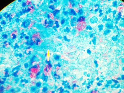
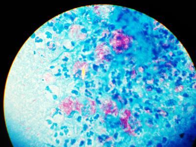
Lymph node aspirates with plentiful AAFB's (red) - Photos: Dr. v. Paleske
However, the microscopy result is often negative for TB-bacilli (AAFBs, Alcohol and Acid fast Bacilli), especially in fairly immune-competent patients, unlike with sputum in cases of TB-lung,.
Helpful in the diagnosis of lymph node TB - despite the absence of microscopically detectable TB-Bacilli - is the detection of caseous material and / or the presence of Langhans giant cells.
The now recommended Xpert MTB/ RIF-test may be quite useful for rapid detection of TB in lymph node aspirates as shown in a study in South Africa with specimens from 50 patients
However, this test cannot replace the need for TB culture to detect MDR- and XDR-TB, as it can only detect Rifampicin- resistance, which alone is not sufficient to qualify for MDR-TB, and the test contributes nothing for the detection of XDR-TB.
Moreover, this test gives a false negative result, if resistance is present not against Rifampicin, but instead against two of the other drugs in use for first line TB-treatment (see Lancet 2011 Vol 377, 1467) .
We introduced the technique of flushing the aspirate needle and syringe with blood taken from the patient and sent it off for TB culture in an EDTA-tube (details here).
We have reported the results of lymph node fine needle aspirates of 156 patients, referred to the Haematology Department at Mpilo-Hospital in Bulawayo /Zimbabwe, during a one year period (Mai 2010 to April 2011) with a high yield of culture positive specimens.
We report here the results of a second study.
Patients and Methods
Lymph node aspirates were performed on 545 patients referred for fine needle aspirate of enlarged peripheral or paraarotic / parailiacal / abdominal lymph nodes during the period from 1st May 2011 to 30th April 2012:.
- 437 patients age 16 – 86, (median age 37 years) 226 female (median age 38years ) and 211 male (median age 35 years)
- 108 patients age 2 months to 15 years, median age 3 years, 58 female (median age 3.83 years) and 50 male (median age 2.83 years)
None of the patients had a formal lymph node biopsy before.
Aspirates were performed with a 5ml or 10 ml syringe. For peripheral lymph nodes size 18 or 19 G needles were used, however for abdominal or paraaortal /parailiacal lymph node aspirates the smaller size 21 G needles.
The specimens were spread on a glass slide and a quash smear with another glass slide facilitated. This technique is better in preserving fragments and thus allows better the recognition of caseous material.
Thereafter a blood sample was taken from the patient and 1 - 1,5ml blood filled in an EDTA-Tube (Full blood count tube).
The aspirate syringe with the aspirate needle on top was flushed several times with the blood in the EDTA-tube by sucking the blood into the aspirate syringe an pressing it back into the EDTA tube, thus delivering the rest of the specimen into the EDTA-tube.
The EDTA tube was then sent to the TB-Lab for TB-culture (solid and liquid media).
May- Gruenwald Giemsa staining was being done on one glass slide, Ziehl-Neelsen for detection of AAFBs on the other.
Results
Altogether the diagnosis TB was made in 232 out of 545 patients (42.56%).
- 25 / 232 (10.77%) in the age group 2 months to 15 years
- 207 / 232 (89.23%) in the age group 16 - 86 years
TB was diagnosed in:
- 23.14 % (25 / 108) of all aspirates in the age group 2 months to 15 years
- 47.36% (207 / 437) in the age group 16 - 86 years
In Detail
1. Microscopically positive for TB (Langhans Giant Cells and / or caseation and / or AAFBs detected) were 215 / 232 (92%), of which 119 / 232 (51%) were microscopically AAFB's-positive
2. TB-Culture positive were 132 / 232 (56%) of all TB diagnoses: 128 mycobacterium tuberculosis, 4 mycobacteria other than m.tuberculosis.
More in Detail:
-- Culture positive, plus microscopically positive including AAFBs detected were 82 / 232 (35.34%)
- Culture positive plus microscopically caseation and / or Langhans giant cells but no AAFBs microscopically detected were 33 / 232 (14.22%)
- Culture positive “only” , microscopically no evidence of TB (neither AAFB's detected, nor caseation, nor Langhans giant cells were found, instead a reactive pattern) was seen in 17 / 232 specimens (7.32% )
- Culture negative but microscopically AAFBs detected were 37 / 232 (15.94%)
- Culture negative, but microscopically positive however no AAFBs detected were 63 / 232 (27.15%)
Patients already on ATT
56 patients had already been started on Anti-TB drugs before they were sent for FNAC, the treatment duration ranging from two days to 6 months. 26 / 56 were culture negative for TB.
Discussion
Compared to our first study we saw a comparatively lower percentage of culture positive TB results: 56% vs. 65,8%. This is to a certain extent possibly the result of aspirates from patients, who had been started on Anti-TB drugs or completed a course at the time of obtaining the specimen.
However looking at the culture results of specimens, which were reported positive for AAFB’s, but negative for TB culture, the figure rose only slightly from 11.3% to 15.94% and may be due to insufficient amount of specimen for culture obtained.
The diagnosis TB was less frequently made in the age group 2 months to 15 years. Many of the patients in this age group, presenting with enlarged lymph nodes, were actually having (Non-TB-) bacterial lymphadenitis.
Conclusions
The aspirate-specimens from lymph nodes mixed with 1-1,5 ml of EDTA-Blood from the patient sent for culture have an acceptable yield, this has been now confirmed with an even much larger number of patients.
Those however, whose aspirates are microscopically AAFBs positive, not on ATT and culture negative are definitely culture false negative and need treatment. A repeat aspirate solely for culture may be appropriate.
In view of the higher number of false negative culture results in this study, a second aspirate for culture in each patient may be advisable.
Patients, whose aspirates are microscopically AAFB´s negative, culture negative, but were diagnosed on the basis of caseation, or pus, or Langhans giant cells, without the presence of Non-TB bacilli, and without a significant amount of granulocytes or macrophages, who are either for weeks on anti-TB-drugs or have recently completed their TB treatment cannot outright be regarded as therapeutic failures. Therefore a change of treatment (switch to 2nd line TB treatment) seems not justified, rather a “wait and see” policy.
 Lymph node aspirates in Tuberculosis-Diagnosis: New challenges, new solutions – a study of 156 patients
 Diagnosis of disseminated Tuberculosis in bone marrow aspirates
References
1) Ligthelm LJ, Nicol MP, Hoek KG, Jacobson R, van Helden PD, Marais BJ, Warren RM, Wright CA:. Xpert MTB/RIF for rapid diagnosis of tuberculous lymphadenitis from fine-needle-aspiration biopsy specimens - J Clin Microbiol. 2011 Nov;49(11):3967-70
2) Pithie AD, Chicksen B.
Fine-needle extrathoracic lymph-node aspiration in HIV-associated sputum-negative tuberculosis, Lancet. 1992 Dec 19-26;340(8834-8835):1504-5
3) Dandapat, M.C. B. M. Mishra, S. P. Dash, P. K. Kar
Peripheral lymph node tuberculosis: A review of 80 cases British Journal of Surgery
Vol. 77, pp. 911–912, 1990
4)Scott K. Heysell, Anthony P. Moll, Neel R. Gandhi, François J. Eksteen, Palav Babaria, Yacoob Coovadia, Lynn Roux, Umesh Lalloo, Gerald Friedland, and N. Sarita Shah
-Extensively Drug-Resistant Mycobacterium tuberculosis from Aspirates, Rural South Africa--- CDC-EID Vol. 16 No 3, March 2010
E-Mail: avonpaleske@yahoo.de
Department of Haematology: Alexander von Paleske, MD, FA(Med), FA (Haem / Onc); Owen Chivima, SCMLT
National TB Reference Laboratory: Barbara Murwira, CMLS; Thembela Masuku, SMLS; Tanaka Sakubani, SMLS; Regina Bhebe, SMLS.
Mpilo-Hospital
Teaching Hospital of the University of Zimbabwe
Bulawayo/Zimbabwe
Africa
Introduction
Lymph node tuberculosis is not infrequent in Sub-Saharan Africa. Even though precise figures do not exist, the incidence can be estimated to be between 10 and 25% of all TB cases.
However:
- 3% of all Tuberculosis cases worldwide are multidrug resistant (MDR-TB). In certain affected areas the percentage is much higher. Thus the diagnosis of MDR-TB is extremely important. The extensive drug resistant TB (XDR-TB) is on the rise as well.
- The HIV epidemic has brought more challenges for the diagnosis of lymph node TB- Many patients present with enlarged lymph nodes of which it is unclear, whether this is TB, or persistent generalized HIV-related lymph node enlargement
- Other infectious diseases like Histoplasmosis and Cryptococcal infections, or simple bacterial lymphadenitis can present with lymph node enlargement as well
- The same applies to HIV-related cancers presenting with lymph node enlargement (Kaposi-Sarcoma, high grade Non Hodgkin lymphoma, Hodgkin´s disease, Castleman's disease).
- HIV-positive patients, who are on anti-TB-treatment (ATT) for lymph node TB, and who have been started concurrently or shortly before on treatment with antiretroviral drugs, can show increasing, or at least persistent lymph node enlargement, which may be in fact not drug-resistant TB, but part of an Immune-reconstitution Syndrome (IRiS), or newly developed high grade lymphoma.
- The new WHO- recommendations for TB-prophylaxis in HIV-positive patients - INH for a period of at least 6 months - require the exclusion of active TB, which is clinically often not possible in view of the frequently enlarged lymph nodes in HIV-positive patients.
The gold standard for a definitive diagnosis is certainly a lymph node biopsy. However this is in most of Sub-Saharan Africa, due to lack of facilities and manpower, at present only feasible for a very small number of patients.
Lymph node aspirates on the other hand are simple to perform, the specimen obtained can easily be spread on slides and then assessed by a laboratory technologist .


Lymph node aspirates with plentiful AAFB's (red) - Photos: Dr. v. Paleske
However, the microscopy result is often negative for TB-bacilli (AAFBs, Alcohol and Acid fast Bacilli), especially in fairly immune-competent patients, unlike with sputum in cases of TB-lung,.
Helpful in the diagnosis of lymph node TB - despite the absence of microscopically detectable TB-Bacilli - is the detection of caseous material and / or the presence of Langhans giant cells.
The now recommended Xpert MTB/ RIF-test may be quite useful for rapid detection of TB in lymph node aspirates as shown in a study in South Africa with specimens from 50 patients
However, this test cannot replace the need for TB culture to detect MDR- and XDR-TB, as it can only detect Rifampicin- resistance, which alone is not sufficient to qualify for MDR-TB, and the test contributes nothing for the detection of XDR-TB.
Moreover, this test gives a false negative result, if resistance is present not against Rifampicin, but instead against two of the other drugs in use for first line TB-treatment (see Lancet 2011 Vol 377, 1467) .
We introduced the technique of flushing the aspirate needle and syringe with blood taken from the patient and sent it off for TB culture in an EDTA-tube (details here).
We have reported the results of lymph node fine needle aspirates of 156 patients, referred to the Haematology Department at Mpilo-Hospital in Bulawayo /Zimbabwe, during a one year period (Mai 2010 to April 2011) with a high yield of culture positive specimens.
We report here the results of a second study.
Patients and Methods
Lymph node aspirates were performed on 545 patients referred for fine needle aspirate of enlarged peripheral or paraarotic / parailiacal / abdominal lymph nodes during the period from 1st May 2011 to 30th April 2012:.
- 437 patients age 16 – 86, (median age 37 years) 226 female (median age 38years ) and 211 male (median age 35 years)
- 108 patients age 2 months to 15 years, median age 3 years, 58 female (median age 3.83 years) and 50 male (median age 2.83 years)
None of the patients had a formal lymph node biopsy before.
Aspirates were performed with a 5ml or 10 ml syringe. For peripheral lymph nodes size 18 or 19 G needles were used, however for abdominal or paraaortal /parailiacal lymph node aspirates the smaller size 21 G needles.
The specimens were spread on a glass slide and a quash smear with another glass slide facilitated. This technique is better in preserving fragments and thus allows better the recognition of caseous material.
Thereafter a blood sample was taken from the patient and 1 - 1,5ml blood filled in an EDTA-Tube (Full blood count tube).
The aspirate syringe with the aspirate needle on top was flushed several times with the blood in the EDTA-tube by sucking the blood into the aspirate syringe an pressing it back into the EDTA tube, thus delivering the rest of the specimen into the EDTA-tube.
The EDTA tube was then sent to the TB-Lab for TB-culture (solid and liquid media).
May- Gruenwald Giemsa staining was being done on one glass slide, Ziehl-Neelsen for detection of AAFBs on the other.
Results
Altogether the diagnosis TB was made in 232 out of 545 patients (42.56%).
- 25 / 232 (10.77%) in the age group 2 months to 15 years
- 207 / 232 (89.23%) in the age group 16 - 86 years
TB was diagnosed in:
- 23.14 % (25 / 108) of all aspirates in the age group 2 months to 15 years
- 47.36% (207 / 437) in the age group 16 - 86 years
In Detail
1. Microscopically positive for TB (Langhans Giant Cells and / or caseation and / or AAFBs detected) were 215 / 232 (92%), of which 119 / 232 (51%) were microscopically AAFB's-positive
2. TB-Culture positive were 132 / 232 (56%) of all TB diagnoses: 128 mycobacterium tuberculosis, 4 mycobacteria other than m.tuberculosis.
More in Detail:
-- Culture positive, plus microscopically positive including AAFBs detected were 82 / 232 (35.34%)
- Culture positive plus microscopically caseation and / or Langhans giant cells but no AAFBs microscopically detected were 33 / 232 (14.22%)
- Culture positive “only” , microscopically no evidence of TB (neither AAFB's detected, nor caseation, nor Langhans giant cells were found, instead a reactive pattern) was seen in 17 / 232 specimens (7.32% )
- Culture negative but microscopically AAFBs detected were 37 / 232 (15.94%)
- Culture negative, but microscopically positive however no AAFBs detected were 63 / 232 (27.15%)
Patients already on ATT
56 patients had already been started on Anti-TB drugs before they were sent for FNAC, the treatment duration ranging from two days to 6 months. 26 / 56 were culture negative for TB.
Discussion
Compared to our first study we saw a comparatively lower percentage of culture positive TB results: 56% vs. 65,8%. This is to a certain extent possibly the result of aspirates from patients, who had been started on Anti-TB drugs or completed a course at the time of obtaining the specimen.
However looking at the culture results of specimens, which were reported positive for AAFB’s, but negative for TB culture, the figure rose only slightly from 11.3% to 15.94% and may be due to insufficient amount of specimen for culture obtained.
The diagnosis TB was less frequently made in the age group 2 months to 15 years. Many of the patients in this age group, presenting with enlarged lymph nodes, were actually having (Non-TB-) bacterial lymphadenitis.
Conclusions
The aspirate-specimens from lymph nodes mixed with 1-1,5 ml of EDTA-Blood from the patient sent for culture have an acceptable yield, this has been now confirmed with an even much larger number of patients.
Those however, whose aspirates are microscopically AAFBs positive, not on ATT and culture negative are definitely culture false negative and need treatment. A repeat aspirate solely for culture may be appropriate.
In view of the higher number of false negative culture results in this study, a second aspirate for culture in each patient may be advisable.
Patients, whose aspirates are microscopically AAFB´s negative, culture negative, but were diagnosed on the basis of caseation, or pus, or Langhans giant cells, without the presence of Non-TB bacilli, and without a significant amount of granulocytes or macrophages, who are either for weeks on anti-TB-drugs or have recently completed their TB treatment cannot outright be regarded as therapeutic failures. Therefore a change of treatment (switch to 2nd line TB treatment) seems not justified, rather a “wait and see” policy.
References
1) Ligthelm LJ, Nicol MP, Hoek KG, Jacobson R, van Helden PD, Marais BJ, Warren RM, Wright CA:. Xpert MTB/RIF for rapid diagnosis of tuberculous lymphadenitis from fine-needle-aspiration biopsy specimens - J Clin Microbiol. 2011 Nov;49(11):3967-70
2) Pithie AD, Chicksen B.
Fine-needle extrathoracic lymph-node aspiration in HIV-associated sputum-negative tuberculosis, Lancet. 1992 Dec 19-26;340(8834-8835):1504-5
3) Dandapat, M.C. B. M. Mishra, S. P. Dash, P. K. Kar
Peripheral lymph node tuberculosis: A review of 80 cases British Journal of Surgery
Vol. 77, pp. 911–912, 1990
4)Scott K. Heysell, Anthony P. Moll, Neel R. Gandhi, François J. Eksteen, Palav Babaria, Yacoob Coovadia, Lynn Roux, Umesh Lalloo, Gerald Friedland, and N. Sarita Shah
-Extensively Drug-Resistant Mycobacterium tuberculosis from Aspirates, Rural South Africa--- CDC-EID Vol. 16 No 3, March 2010
E-Mail: avonpaleske@yahoo.de
onlinedienst - 15. Aug, 21:29 Article 11837x read
